The browser you are using is not supported by this website. All versions of Internet Explorer are no longer supported, either by us or Microsoft (read more here: https://www.microsoft.com/en-us/microsoft-365/windows/end-of-ie-support).

Please use a modern browser to fully experience our website, such as the newest versions of Edge, Chrome, Firefox or Safari etc.

A method for measuring the healing after surgery

White background and different red blobs in the foreground. The word cancer is written on the red blob that corresponds to the stomach. Illustration.
Esophagus cancer is treated with surgery by stitching the upper stomach together with the healthy part of the esophagus. An optical fiber can send light down the esophagus to look for problems occurring after the surgery.

Imagine you had to get surgery due to esophagus cancer. Initially, you seem to recover well, but suddenly you are rushed to emergency surgery. The wound has failed to heal and the inflammation has caused severe, life-threatening sepsis. What if this could have been prevented? What if your doctors could have seen the problem in the wound days before the severe problem occurred, and been able to fix it before it almost killed you? This project will develop a method that can be used to measure how well the surgery scar after gastric surgery is healing, and help doctors discover problems with recovery after gastric surgery before the problem becomes life-threatening.

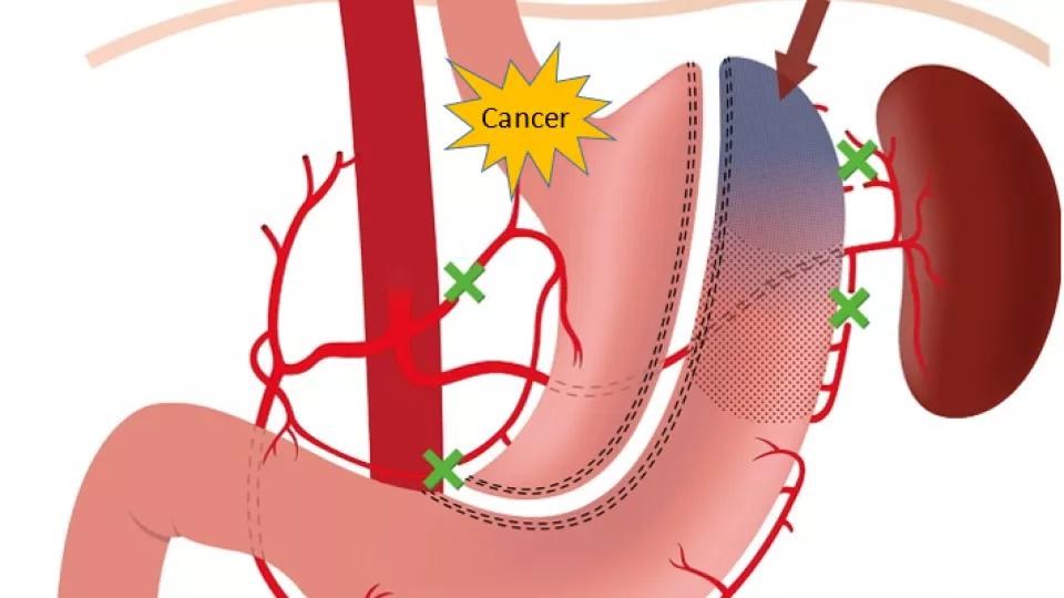
Cancer in the esophagus is one of the most increasing types of cancer in the western world, and often surgery is the only treatment. In this treatment, part of the esophagus is removed, and the upper stomach is reconstructed to splice together with the remaining esophagus. In 15-25% of all patients, the joint fails to heal well, leading to severe inflammation and/or sepsis, which are difficult to fix and potentially life-threatening.

In this project, we will study the blood circulation around the joint after esophageal surgery to see if we can detect problems with healing earlier. We will add a substance to the blood that fluoresces (=starts to glow) when illuminated by a certain type of infrared light. By illuminating the joint with infrared light through an optic fiber and then photographing an image of the so-called fluorescence light from the injected substance, indocyanine green, in the blood, we hope to be able to measure and early identify problems in the blood circulation that may be the first sign of problems in the healing of the joint.

This will help doctors identify and fix problems with healing after gastric surgery at an early stage, which will improve the survival rate and recovery of patients with esophagus cancer.

Anna-Lena Sahlberg - portal.research.lu.se